Congratulations!
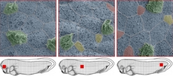
Using electron micrographs, it becomes evident that the cellular composition of the frog embryonic skin is varied in different areas. The skin of frog embryos is very similar to the human airway epithelium. It contains mucus secretory cells (red and blue) as well as ciliated cells (green). Together, these cell types clean the airways and protect from infections. This process only works correctly when the right amount of correct cell types is combined at different locations. Dr. Martin Helmstädter (EM) and Dr. Peter Walentek
Scientists from Freiburg use the embryonic skin of frogs to understand how lung tissues are organized, because they both have common features. The goal is to understand how the right cells are formed in the right place.
How the human lung develops and which processes are altered in disease remains incompletely understood. Therefore, scientists from the Medical Faculty of the Freiburg University are investigating the organization processes, which occur during development of the embryonic epidermis of frog tadpoles. Similar to the lung and airways, the skin of tadpoles is composed of cells that release mucus as well as cells that form tiny hair-like structures on their surface to move fluid over the epithelium to clean it. The scientists from the Clinic for Internal Medicine IV of the University Freiburg Medical Center, together with the French team of Dr. Laurent Kodjabachian, could now win a highly competitive grant from the French Agence nationale de la recherche (ANR) and the German DFG to fund a bilateral project over the next 3 years with over 550.000 €.
“We want to establish a cellular atlas of the embryonic frog skin. This atlas will also allow important insights into how airway epithelia are formed” says Dr. Peter Walentek, group leader at the Clinic for Internal Medicine IV of the University Freiburg Medical Center. “When we gain a better understanding in the compositions and functions of different cell types, it will also help us to understand better how and why lung tissues change during human disease, a prerequisite to develop new therapeutic strategies in the future” says Walentek. This is because in chronic airway diseases, changes in the composition of cell types are frequently observed, which leads to a failure in airway clearance. The patients then have a strongly increase risk for lung infections.
Learning from the African clawed frog Xenopus about human disease.
Humans and Xenopus are both vertebrates and they share about 21.000 of 23.000 genes. This allows scientists to study aspects of human development and disease using the frog embryo. This also helps to partially replace studies using mammalian systems. Thus, this strategy helps to implement the 3-R principles of animal research, reducing the number of research animals used (Reduce), improving experimental strategies (Refine), or using simpler animals for studies (Replace). Another advantage is: “The animals lay a huge amount of eggs and embryos develop in a petri dish, which makes it very easy to manipulate them or to observe them by light microscopy. Furthermore, it is easy and fast to manipulate gene expression and functions in order to study the gene’s contributions to development and disease”, according to Walentek.
Text by: J. Faber